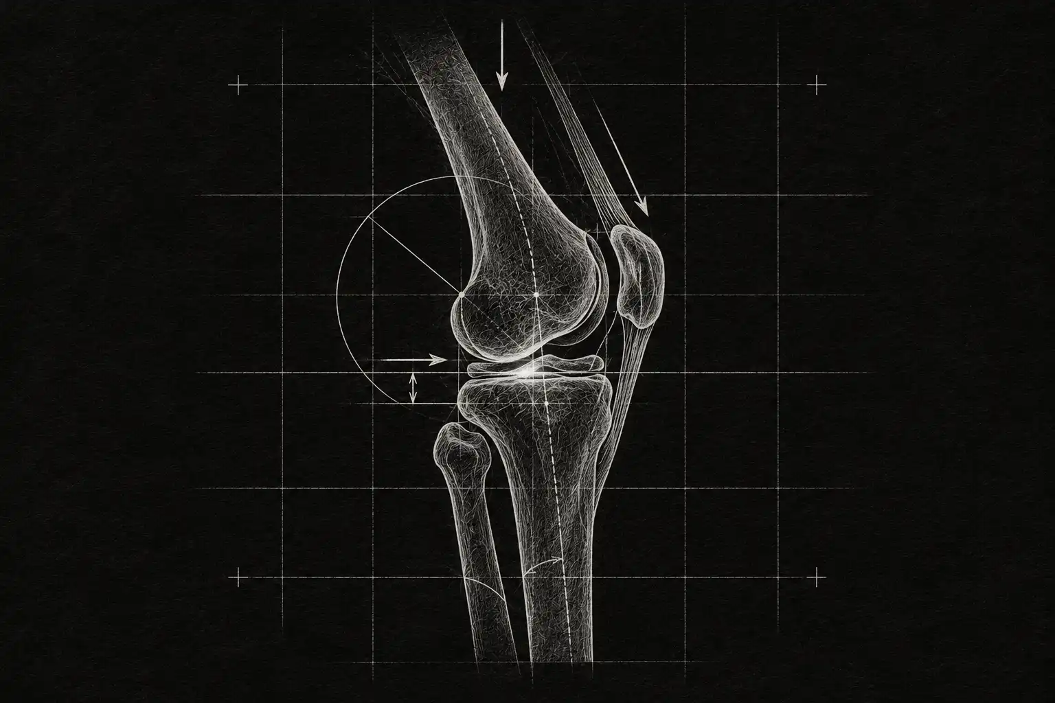
Your knee hurts on the stairs or when you get up from a chair, and you’ve been told you have “osteoarthritis”: “worn out”, “bone on bone”, “better not run”, “you’ll need surgery”. This guide separates what we know from what gets repeated without evidence.
Looking for what to do? Go straight to the recovery plan
What knee osteoarthritis is
Knee osteoarthritis is a chronic joint problem, with activity-related pain and, sometimes, brief morning stiffness.[2] It doesn’t mean “wear and tear” or “the end of movement”: pain doesn’t depend only on what shows on a scan.[3]
See more
It usually appears after age 40. There is currently no treatment that cures it,[8] but the guidelines agree that exercise, weight management and information help people live better.[2]
Knee anatomy: your joint, in layers
Tap each layer (or the figure itself) to see what it is, what happens to it when it hurts… and above all, what it doesn’t mean.
Bones: the structure that holds you up
What it is. The femur above, the tibia below and the kneecap in front, sliding over the femur. They spread the body’s weight and give the muscles leverage.
When it hurts. With age, osteophytes (small rims of new bone) and changes in the bone under the cartilage appear. They are very common: they show up in three out of four knees of people over 50 who have no osteoarthritis on X-ray.[1]
Careful. It doesn’t mean: “I have osteophytes, that’s why it hurts”. They are a very common finding and imaging alone does not explain the pain.[9]
Cartilage: the sliding surface
What it is. A smooth, slippery layer covering the ends of the femur and tibia and the back of the kneecap. It reduces friction and spreads the load.
When it hurts. In osteoarthritis the cartilage thins and changes, but the disease is not limited to cartilage: it also involves the bone, the synovial lining, the ligaments and the muscles.[10]
Careful. It doesn’t mean: “I have no cartilage left, nothing can be done”. Cartilage damage shows up in 7 out of 10 knees of people over 50 with no osteoarthritis on X-ray.[1]
Menisci: the shock absorbers
What it is. Two crescent-shaped pads of fibrocartilage between the femur and the tibia. They cushion and stabilise.
When it hurts. With age, degenerative meniscal lesions can appear. Arthroscopic surgery for them has not shown an important benefit compared with sham surgery.[11]
Careful. It doesn’t mean: “I have a torn meniscus, I need surgery”.
Ligaments and capsule: the stabilisers
What it is. The cruciate ligaments (inside) and the collateral ligaments (on the sides) stabilise the knee. The capsule, lined by the synovial membrane, wraps the joint and produces the fluid that lubricates it.
When it hurts. Inflammation of the synovial lining (synovitis) is common in osteoarthritis,[10] and stiffness appears after a period of inactivity.[2]
Careful. It doesn’t mean: “my knee is unstable and will give way” just because you feel stiffness or hear clicks when you move it.
Muscles: a team, not a soloist
What it is. Quadriceps in front, hamstrings behind, with glutes and calves as partners. They absorb impact and hold the knee up with every step.
When it hurts. People with osteoarthritis tend to be more sedentary.[10] Greater muscle strength, better mental health and more aerobic exercise were linked to a better course in the first three years.[12]
Careful. It doesn’t mean: “my muscles are weak, that’s why I have osteoarthritis”. Strength can be trained, and training it is part of the treatment.[2]
What a knee with osteoarthritis feels like: the gears picture
Many people describe their knee as a gear that no longer turns smoothly. It helps explain what you notice, with a warning at the end. Switch state and watch how the knee bends and straightens.
No osteoarthritis
The parts fit and the movement is smooth: climbing stairs or getting up from a chair is barely noticed.
With osteoarthritis
Movement loses smoothness: activity-related pain and, sometimes, brief morning stiffness.[2] Some people describe it as something that “catches” or creaks when they start moving.
Illustration: the movement is a simulation, not a measurement. The “dirt” only stands for movement becoming rough; osteoarthritis is not dirt.
But a knee is not a machine that simply wears out. Changes on imaging are very common in knees without pain too,[1] and exercise is one of the pillars of treatment, because it helps improve pain and function.[2] The gears describe what you feel, not the fate of the knee.
What your scan does (and doesn’t) say
If you’ve had an X-ray or an MRI, the report may say “osteophytes”, “cartilage damage” or “meniscal lesion”: it sounds serious. See what happens in people over 50 whose X-ray shows no osteoarthritis.
| Imaging finding | On MRI |
|---|---|
| Any abnormality | 89 % |
| Osteophytes | 74 % |
| Cartilage damage | 69 % |
| Bone marrow lesions | 52 % |
Data from 710 people in the Framingham cohort (mean age 62); 29% had knee pain in the past month.[1]
X-ray and pain only partly match:[9] that’s why knee osteoarthritis is diagnosed from the story and the examination, without imaging, from age 45 with activity-related pain and morning stiffness under 30 minutes.[2]
See more
X-ray is not a good guide to pain either: among people with knee pain, the proportion with osteoarthritis on X-ray ranges from 15 to 76%, and among people with osteoarthritis on X-ray, the proportion with pain ranges from 15 to 81%.[9] That’s why imaging should not be used on its own to assess a knee.
And that’s why knee osteoarthritis is diagnosed from the story and the examination: from age 45, with activity-related pain and either no morning stiffness or stiffness lasting under 30 minutes, no imaging is needed to confirm it.[2]
How knee osteoarthritis changes over time
It’s often assumed that osteoarthritis “only gets worse”. Follow-up studies tell another story: this is how pain evolved in the mid-term in seven studies with 7,747 people. Compare the two ideas and tap each channel.
Stays the same: 85 out of 100
The great majority keep a similar level of pain throughout follow-up, with better and worse days. It is not a downhill slope.
Improves: 8 out of 100
A small share notice less pain over time.
Worsens: 7 out of 100
A small share get worse. In the studies, having less education, other illnesses or depression was associated with a worse course.[3]
Percentage of patients by pain trajectory in the mid-term, in seven studies with 7,747 people (systematic review).[3] In almost half, daily pain is unstable: it changes from one day to the next.
These are data from many people, not your own path: yours may vary from one day to the next, and that’s normal.
See more
[12] In another study of 705 people with early osteoarthritis, a higher BMI, other illnesses and more activity limitation were associated with more pain, and severity on X-ray was not.[13]
What makes your pain go well or badly?
Pain doesn’t depend only on what shows on a scan. These are the factors that, in follow-up studies, go with a better or a worse course. Tap each one.
Weight: a higher BMI
In a study of 705 people with early osteoarthritis, a higher BMI was associated with more pain and with worsening pain.[13] The other side: in overweight, combining diet and exercise reduced pain moderately.[14]
Other illnesses
Having other illnesses was associated with a worse course of pain, both in a study of 705 people[13] and in a review of seven studies.[3]
Mood: depression
Depression was associated with more severe or worsening pain;[3] and better mental health, with a better course in the first three years (in a review of hip or knee osteoarthritis).[12]
Level of education
Having less education was associated with a worse course. It is not something that changes in the clinic, but it helps to think about who needs more support and a clearer explanation.[3]
Muscle strength and aerobic exercise
Greater muscle strength and more aerobic exercise were associated with a better course in the first three years (hip or knee osteoarthritis).[12]
Social support and self-confidence
Social support and self-efficacy (confidence in being able to manage the pain) also appear among the protective factors of that review.[12]
Severity on X-ray
Neither the review of seven studies[3] nor the study of 705 people[13] found a clear relationship between baseline radiographic severity and how the pain evolved.
These are associations seen in follow-up studies: they do not prove the factor is the cause, and they can’t predict what will happen to one particular person.
The good news: much of it can be worked on. Train strength, look after weight if there is excess weight, and seek support if mood is low. In a trial of 454 people with overweight, losing 11% of body weight with diet and exercise gave less pain and better function than diet alone or exercise alone.[15]
Knee osteoarthritis myths: five ideas worth letting go of
Tap each card to turn it over and see what the evidence says.
“My knee is worn out: bone on bone.”
The changes seen on imaging are very common: in people over 50 with no osteoarthritis on X-ray, MRI shows some abnormality in 89%, with or without pain.[1] And X-ray and pain only partly match.[9] A knee with changes isn’t a knee that inevitably has to hurt.
“Running ruins your knees.”
In a meta-analysis of 25 studies (125,810 people), hip or knee osteoarthritis was present in 3.5% of recreational runners, compared with 10.2% of sedentary people and 13.3% of competitive runners.[16] Recreational running did not appear as a risk (versus sedentary people, OR 0.86; 95% CI 0.69–1.07, not statistically significant). This figure is about who develops osteoarthritis; if you already have it, what matters is adapting the load with your physiotherapist.
“I need an MRI to know what’s wrong.”
Knee osteoarthritis is diagnosed from the story and the examination, without imaging, from age 45 if there is activity-related pain and morning stiffness is absent or lasts under 30 minutes. Guidelines don’t recommend routine imaging to diagnose it or to follow it up, unless there are atypical features.[2]
“An arthroscopy will clean up my knee.”
Arthroscopic surgery (washout, debridement or trimming of the meniscus) in knee osteoarthritis and degenerative meniscal tears does not importantly improve pain or function compared with sham surgery, with high-certainty evidence.[11] That’s why NICE recommends not offering arthroscopic lavage or debridement.[2]
“An injection will regenerate my cartilage.”
Corticosteroid injections give short-lived relief (2 to 10 weeks, according to NICE).[2] In a 2-year trial of 140 people, corticosteroid injections led to more cartilage loss than saline and did not reduce pain more.[17] And NICE does not recommend hyaluronic acid injections.[2]
Knee osteoarthritis treatment: what actually helps
Not every treatment has the same backing. This is how the international guidelines rank them:[2][18][19][20] from the most recommended to what is better avoided. Tap each one.
Rung 1 · Foundations: Education, exercise and weight
Tags: Information and support · Therapeutic exercise · Weight management
- NICE recommends offering everyone with osteoarthritis therapeutic exercise tailored to their needs (local strengthening, aerobic fitness) and considering combining it with education or behaviour change.[2]
- OARSI places education and land-based exercise programmes, with or without diet management, as the core treatments for the knee.[18] The American College of Rheumatology backs this with strong recommendations for exercise, weight loss in people with overweight and self-management programmes.[19]
- If there is excess weight, NICE explains that any weight loss is likely to be beneficial and that losing 10% of body weight is probably better than losing 5%.[2]
Rung 2 · Add-ons: Occasional, symptom-focused support
Tags: Topical NSAIDs · Oral NSAIDs, with caution · Walking stick · Occasional corticosteroid
- NICE recommends offering a topical anti-inflammatory for the knee; if that isn’t enough, considering an oral one with stomach protection and taking the risks into account.[2] OARSI advises against oral anti-inflammatories in people with cardiovascular disease or frailty.[18]
- A meta-analysis of 76 trials with more than 58,000 patients found that all the anti-inflammatories studied improved pain on average compared with placebo, although only some (such as high-dose diclofenac or etoricoxib) reached a clinically important improvement.[21]
- NICE suggests considering a walking stick in lower-limb osteoarthritis.[2] And it considers a corticosteroid injection when everything else fails or to support exercise, warning that the relief lasts 2 to 10 weeks.[2]
- They work best as part of an overall plan: they ease symptoms to make movement easier. Any drug should be assessed by your doctor.
Rung 3 · Use with caution: Little support, or risks that outweigh benefits
Tags: Hyaluronic acid · Arthroscopy · Glucosamine and opioids · Paracetamol
- NICE recommends not offering hyaluronic acid injections or arthroscopic lavage or debridement.[2] The guidelines don’t fully agree: OARSI keeps hyaluronic acid as a second-level option depending on the person’s other conditions.[18]
- NICE advises against glucosamine and strong opioids, and does not routinely recommend paracetamol or weak opioids.[2] In a meta-analysis, paracetamol in hip or knee osteoarthritis had a significant but not clinically important effect on pain.[22]
- They also differ on acupuncture: NICE doesn’t offer it,[2] and the American College of Rheumatology recommends it conditionally.[19]
How much does exercise help, and will you notice?
The most recent Cochrane review (139 trials, 12,468 people) finds that exercise improves pain, function and quality of life. Is that improvement big enough to notice? Pick an outcome and see.
Pain
Exercise versus placebo or attention control. 8.7 (5.7 to 11.7). Certainty: low.
Exercise versus doing nothing or usual care. 13.1 (10.4 to 15.9). Certainty: low.
Exercise added to another treatment. 10.4 (8.1 to 12.8). Certainty: moderate.
Function
Exercise versus placebo or attention control. 11.3 (7.6 to 15.1). Certainty: moderate.
Exercise versus doing nothing or usual care. 12.5 (9.7 to 15.3). Certainty: moderate.
Exercise added to another treatment. 9.7 (7.5 to 12.0). Certainty: moderate.
Quality of life
Exercise versus placebo or attention control. 6.1 (-0.1 to 12.3). Certainty: moderate.
Exercise versus doing nothing or usual care. 5.4 (3.2 to 7.5). Certainty: moderate.
Exercise added to another treatment. 4.2 (1.4 to 7.1). Certainty: moderate.
Source: 2024 Cochrane review (0 to 100 scale, right after the intervention).[23]
What to take away: exercise is the foundation of treatment in the guidelines[2]; paracetamol barely helps[22] and arthroscopic clean-up does no better than a simulated operation.[11]
See more
What counts is consistency and what you value: climbing stairs, walking without pain.[2] The minimal important difference is the change a person usually really notices: 12 points for pain, 13 for function and 15 for quality of life, out of 100.
Recovery plan in phases: exercise and movement
Everyone is different, but the path tends to follow this order. These are examples to understand the logic, not a personal prescription.
Phase 1 · Understand and settle (≈ weeks 0–2)
Goal. Turn the alarm down: understand what’s going on, reduce fear and keep moving without forcing it. It’s normal for pain to go up a little when you start exercising; consistency is what brings the benefit.[2]
What you can do:
- Carry on with daily life, adapting loads rather than stopping
- Walk at a comfortable pace, in short bouts
- Change position often
Phase 2 · Move with confidence (≈ weeks 2–8)
Goal. Regain range of movement and return to the actions you were avoiding out of fear.
What you can do:
- Gentle mobility within comfort
- Progressive walking; stairs and sitting down and standing up from a chair in sets
- Gradually return to what you avoid (squatting, kneeling, climbing stairs)
Phase 3 · Build capacity (≈ weeks 8–16)
Goal. Gain strength and load tolerance so your knee can take more.
What you can do:
- Progressive quadriceps, glute and calf strength
- Use everyday loads (shopping, gardening, kids) as training
- Aerobic exercise (brisk walking, cycling, swimming) to taste
Phase 4 · Maintain and prevent flare-ups (≈ from week 16)
Goal. Make it a habit: keeping exercise up in the long term increases its benefits.[2] And have a plan for bad days.
What you can do:
- A realistic exercise routine: the one you can keep up
- A “bad day” plan: what you reduce and what you keep
- Check in on weight, sleep and work from time to time
The full map: body, mind and surroundings
Persistent pain almost never has a single cause: body, mind and surroundings add up. That’s why it helps to look at all three at once.
What I see in clinic
With the knee I see something similar to what I see with the back: what repeats most is not the knee, it’s the sentence someone said to them.
They arrive with an X-ray or an MRI in hand and one fixed idea: that the knee is “worn out”, that it’s “bone on bone” and that it’s best not to push it. Many have already given up stairs, walks or sport for fear of making it worse.
What tends to change things is not a miracle technique. It’s understanding what that image really says, going back to moving with a load the knee tolerates, and giving strength time.
Osteoarthritis doesn’t go away, but once you stop seeing it as a machine that breaks, it stops deciding what you do with your life.
For professionals and students
Show the technical contentEvidence tables, referral criteria and clinical practice guidelines, in technical terms.
The evidence and clinical reasoning behind the article, in technical terms. Figures come from each source’s official abstract; our own calculations are flagged. Tap an underlined abbreviation to see what it means.
Abbreviations
RCT randomised controlled trial · CI confidence interval · MD mean difference · SMD standardised mean difference · KOOS Knee injury and Osteoarthritis Outcome Score · NSAID non-steroidal anti-inflammatory drug · MRI magnetic resonance imaging · BMI body mass index · VAS visual analogue scale for pain · NRS numeric rating scale for pain (0–10) · WOMAC Western Ontario and McMaster Universities Osteoarthritis Index · PHQ-9 Patient Health Questionnaire-9, depression questionnaire · PSEQ Pain Self-Efficacy Questionnaire · ACR American College of Rheumatology · HIV human immunodeficiency virus · OARSI Osteoarthritis Research Society International · EULAR European Alliance of Associations for Rheumatology · NICE National Institute for Health and Care Excellence, the UK public body for clinical guidelines
Clinical assessment and prognosis
Key points: clinical diagnosis without imaging (from age 45, activity-related pain and morning stiffness of up to 30 minutes); rule out what needs another route; measure pain and function with the KOOS and performance tests at every review.
Clinical pathway
- DiagnoseClinical diagnosis without imaging if the person is 45 or over, has activity-related pain and morning stiffness that is absent or lasts up to 30 minutes.[2]
- ScreenRule out what needs another route: a hot joint with fever, trauma with inability to bear weight, mechanical locking, signs of thrombosis (see “Refer”).
- StratifyIdentify what is associated with a worse course: high BMI, other illnesses and greater activity limitation;[13] depression, in another review.[3]
- TreatEducation, exercise and weight management as the base (see “Treat”).
- MeasurePain and function with a questionnaire (KOOS)[24] and performance tests (chair stand, walking, stairs)[25] at baseline and at each review.
- ReassessNo improvement with an adequate conservative approach: review the diagnosis and consider referral for joint replacement assessment (see “Refer”).
Before measuring
- Imaging
- Not routinely, neither to diagnose nor to follow up, unless there are atypical features or features suggesting another diagnosis.[2]
- Septic arthritis
- In a painful, swollen joint, look for risk factors: age, diabetes, rheumatoid arthritis, previous joint surgery, hip or knee prosthesis, skin infection or HIV. Joint pain (sensitivity 85%), swelling (78%) and fever (57%) were the only findings present in more than half of the cases: fever may be absent.[7]
Minimum set: at every visit
- Scale
- 5 subscales · 0–100
- Relevant change
- Minimal detectable change ≥ 20 in older adults[24]
Questionnaire with subscales for pain, symptoms, daily living, sport and quality of life. The daily-living subscale fits older people better, and the pain subscale fits painful knees.[24]
- Scale
- 3 core tests
30-second chair stand, 40 m fast-paced walk and stair climb. They complement questionnaires.[25]
- Scale
- 0–10 · one question
Rating of pain intensity from 0 (no pain) to 10 (worst imaginable). Its measurement properties were reviewed alongside the VAS and other adult pain scales.[26]
Use it when you need a quick pain measure at every visit.
- Scale
- 3 subscales: pain, stiffness and function
Self-administered questionnaire for hip or knee osteoarthritis. In its validation study the pain, stiffness and function subscales met the criteria for validity, reliability and responsiveness.[27]
Use it when you prefer a questionnaire specific to knee or hip osteoarthritis, as an alternative to the KOOS.
According to what you find
- Scale
- 9 items · 0–27
Brief depression severity questionnaire: each item scores 0 to 3, and 5, 10, 15 and 20 points mean mild, moderate, moderately severe and severe depression. With 10 or more, sensitivity and specificity for major depression were 88%.[28]
Use it when mood is low or the pain is not progressing as expected: depression was associated with a worse course.[3]
It measures confidence in doing activities despite the pain, not only in controlling it.[29]
Use it when you suspect a lack of confidence to move: self-efficacy appears among the protective factors.[12]
Classification criteria developed for research: they use algorithms with history, examination, laboratory tests and radiographs, and compared osteoarthritis with rheumatoid arthritis and other knee pain.[30]
Use it when you need to classify knee osteoarthritis for research or read studies that use them.
What to look for when assessing
- What they have stopped doing
- Ask which activities they have given up because of the pain and why: the pain itself, fear, or advice they were given?
- What they think is wrong
- Explore how they explain their problem: “bone on bone” or “wear and tear” often shape how much they move.
- The same measure from day one
- Record the starting value of the questionnaire and the tests at the first visit and always use the same version, so that reviews can be compared with each other.
Treatment: certainty of evidence
Key points: exercise improves pain and function (low to moderate certainty); arthroscopy does no better than sham surgery (high certainty); sustained corticosteroid did not reduce pain more and was associated with more cartilage loss.
Certainty of the evidence:HighModerateLow
| Intervention | What it means | Certainty | Key data |
|---|---|---|---|
| Exercise versus no treatment, usual care or limited education | Improves pain and function, but the gain may fall short of the minimal important difference. | Low | −13.1MD in pain (0–100) 95% CI −15.9 to −10.4[23] Function: −12.5 (−15.3 to −9.7), moderate certainty 56 studies · n = 4,184 |
| Exercise versus placebo or attention control | Improves pain a little; part of the effect may come from the participant knowing they are being treated. | Low | −8.7MD in pain (0–100) 95% CI −11.7 to −5.7 Quality of life: no clear improvement 28 studies · n = 2,873 |
| Exercise added to another intervention | Adds to what is already being done (diet, physiotherapy, education). | Moderate | −10.4MD in pain (0–100) 95% CI −12.8 to −8.1 47 studies · n = 4,441 · same review |
| Diet + exercise in overweight or obesity | Reduces pain moderately; diet alone did not. | — | −0.37SMD in pain 95% CI −0.69 to −0.04[14] Diet alone: −0.13 (−0.37 to 0.10), not significant meta-analysis of 9 RCTs |
| Arthroscopy versus sham surgery | Does not improve pain or function importantly. | High | −4.6MD in pain at 3 months (0–100) 95% CI −9.0 to −0.02[11] Function: 0.1 (−3.2 to 3.4) 4 RCTs · n = 309 |
| Total knee replacement versus non-surgical treatment (12 months) | Improves pain and function more, with more serious adverse events. | — | 15.8MD in KOOS4 (0–100) 95% CI 10.0 to 21.5[31] Serious adverse events: 24 vs 6 1 RCT · n = 95 who completed follow-up |
| Intra-articular corticosteroid for 2 years versus saline | Did not reduce pain more and was associated with more cartilage loss. | — | −0.11MD in cartilage thickness (mm) 95% CI −0.20 to −0.03[17] Pain: no difference (−0.6; −1.6 to 0.3) 1 RCT · n = 140 |
Certainty as rated by each source; “—” means it is not stated in the abstract consulted. For exercise and arthroscopy, negative values indicate improvement.
| Variable | What the evidence says |
|---|---|
| Type of exercise | Aerobic, resistance and performance exercise reduced pain similarly (SMD 0.67, 0.62 and 0.48), and single-type programmes outperformed mixed ones (0.61 versus 0.16).[32] The 2024 Cochrane review found no differences between types,[23] whereas a network meta-analysis of 217 RCTs placed aerobic exercise as the most likely to be the best.[33] |
| Target muscle | More pain reduction with quadriceps-specific exercise than with general lower-limb exercise (SMD 0.85 versus 0.39).[32] |
| Frequency | With supervised exercise, at least 3 times a week reduced pain more (SMD 0.68 versus 0.41).[32] The 2024 Cochrane review found no relationship between the number of sessions and improvement.[23] |
| Intensity and session length | No effect on pain in the meta-regression.[32] |
| Dose and progression | EULAR asks for dose and progression to be tailored to each person.[20] |
| Long term | NICE states that keeping exercise up in the long term increases its benefits.[2] |
The reviews do not fully agree: use this as guidance, not as a fixed prescription. In the 2014 meta-analysis (SMD), higher values mean greater pain reduction.
| Better to avoid | Better to say |
|---|---|
| “Your knee is worn out: bone on bone.” | “The changes on the scan are common and don’t explain the pain on their own; what matters is how it moves and what we do.” |
| “If it hurts, don’t exercise.” | “It’s normal for pain to go up a little at the start; consistency is what helps.” |
| “This only gets worse and in the end you’ll need surgery.” | “In most people the pain stays stable; a replacement is considered if everything else doesn’t work.” |
Illustrative phrases, written as examples.
Referral criteria
Key points: suspected septic arthritis = urgent medical assessment; trauma with inability to bear weight, suspected deep vein thrombosis or mechanical locking = prioritised assessment; no improvement with conservative treatment = consider joint replacement.
Assess with a clinical rule (Ottawa knee rule) to decide whether an X-ray is needed: in adults it has a sensitivity of 98.5% to rule out fracture.[5]
Swollen, warm, painful calf in one leg only: prioritised medical assessment.
Prioritised specialist assessment, without waiting for conservative treatment to run its course.
NICE suggests considering referral for joint replacement if symptoms substantially affect quality of life and non-surgical management is ineffective or unsuitable; do not exclude by age or use numerical severity scores.[2] In a trial, joint replacement improved more than non-surgical treatment at 12 months, with more serious adverse events.[31]
Official guidelines and documents
Key points: NICE, OARSI, the American College of Rheumatology with the Arthritis Foundation and EULAR agree that exercise, weight management and information are the foundation.
Clinical diagnosis without imaging, therapeutic exercise for everyone, weight management, topical anti-inflammatory for the knee, and not offering hyaluronic acid or arthroscopic lavage or debridement.[2]
Open the original documentnice.org.ukCore treatments for the knee: education and land-based exercise, with or without diet management. Topical anti-inflammatories strongly recommended; paracetamol conditionally not recommended; opioids strongly recommended against.[18]
Open the original documentdoi.orgStrong recommendations for exercise, weight loss, self-management programmes, tai chi, cane use, topical and oral NSAIDs and intra-articular glucocorticoids in knee osteoarthritis.[19]
Open the original documentdoi.orgEight non-pharmacological recommendations: individualised multicomponent plan, information and self-management, exercise with tailored dose and progression, mode of delivery, weight, footwear and walking aids, work-related advice and behaviour change techniques.[20]
Open the original documentdoi.orgKeep exploring
What if your situation doesn’t quite fit this guide? A few pointers to tell the difference:
 PhysiotherapyAug 24, 2026 Every day you don’t move, your body thinks you’re dying Read more
 PhysiotherapyAug 16, 2026 What is pain? Your body’s alarm system Read more
Frequently asked questions about knee osteoarthritis
Does knee osteoarthritis go away?
There is currently no treatment that cures it,[8] but many people keep a stable level of pain[3] and improve with exercise, weight management and information.[2]
Does walking or running make knee osteoarthritis worse?
Exercise is part of the treatment: it may hurt a little more at the start.[2] Recreational running did not appear as a risk.[16]
See more
Exercise is part of the treatment: guidelines recommend offering it to everyone with osteoarthritis and warn that it may hurt a little more at the start.[2] As for running, in a meta-analysis osteoarthritis was less common in recreational runners (3.5%) than in sedentary people (10.2%).[16] If you already have osteoarthritis, the key is the dose: start gradually and adapt the load with your physiotherapist.
Do injections help?
They relieve pain only briefly: 2 to 10 weeks with corticosteroid.[2] Maintained for two years, they did not reduce pain more and were associated with more cartilage loss.[17]
See more
They relieve pain only briefly. Corticosteroid ones can relieve pain for 2 to 10 weeks, according to NICE, which considers them when everything else fails.[2] Maintained for two years, in one trial they did not reduce pain more and were associated with more cartilage loss.[17] NICE does not recommend hyaluronic acid ones.[2] Any injection should be assessed by your doctor.
When is surgery needed?
When everything else hasn’t worked and symptoms substantially affect quality of life.[2] Arthroscopic clean-up is not recommended.[2]
See more
When everything else hasn’t worked. NICE suggests referral for a joint replacement if symptoms substantially affect quality of life and non-surgical management doesn’t work or isn’t suitable.[2] In one trial, joint replacement improved more than non-surgical treatment at 12 months, with more serious adverse events, and only 26% of the non-surgical group had surgery in that time.[31] Arthroscopic clean-up is not recommended.[2]
Does this guide replace a professional assessment?
No. It is educational: if you have warning signs or the pain limits you a lot, see a licensed health professional.
See more
No. Its purpose is educational. If you have warning signs or the pain limits you significantly, see a licensed health professional for an individual assessment.
Want us to look at it together?
Understanding what’s going on in your knee is the first step. The next is designing, with you, a plan that fits your body, your work and your life.
Our contactReferences
Show the 33 references
- [1]Guermazi A, Niu J, Hayashi D, Roemer FW, Englund M, Neogi T, et al. Prevalence of abnormalities in knees detected by MRI in adults without knee osteoarthritis: population based observational study (Framingham Osteoarthritis Study). BMJ. 2012;345:e5339.Observational study↗
- [2]National Institute for Health and Care Excellence. Osteoarthritis in over 16s: diagnosis and management (NG226). Published 19 October 2022.Guideline↗
- [3]Previtali D, Andriolo L, Di Laura Frattura G, Boffa A, Candrian C, Zaffagnini S, et al. Pain Trajectories in Knee Osteoarthritis-A Systematic Review and Best Evidence Synthesis on Pain Predictors. J Clin Med. 2020;9(9):E2828.Systematic review↗
- [4]NHS. Knee pain: when to get medical help. nhs.uk (page last reviewed 21 December 2023; consulted 7 November 2026).Official health source↗
- [5]Bachmann LM, Haberzeth S, Steurer J, ter Riet G. The accuracy of the Ottawa knee rule to rule out knee fractures: a systematic review. Ann Intern Med. 2004;140(2):121-124.Systematic review↗
- [6]NHS. Deep vein thrombosis (DVT): symptoms. nhs.uk (consulted 7 November 2026).Official health source↗
- [7]Margaretten ME, Kohlwes J, Moore D, Bent S. Does this adult patient have septic arthritis? JAMA. 2007;297(13):1478-1488.Systematic review↗
- [8]GBD 2021 Osteoarthritis Collaborators. Global, regional, and national burden of osteoarthritis, 1990-2020 and projections to 2050: a systematic analysis for the Global Burden of Disease Study 2021. Lancet Rheumatol. 2023;5(9):e508-e522.Review↗
- [9]Bedson J, Croft PR. The discordance between clinical and radiographic knee osteoarthritis: a systematic search and summary of the literature. BMC Musculoskelet Disord. 2008;9:116.Systematic review↗
- [10]Katz JN, Arant KR, Loeser RF. Diagnosis and Treatment of Hip and Knee Osteoarthritis: A Review. JAMA. 2021;325(6):568-578.Review↗
- [11]O'Connor D, Johnston RV, Brignardello-Petersen R, Poolman RW, Cyril S, Vandvik PO, et al. Arthroscopic surgery for degenerative knee disease (osteoarthritis including degenerative meniscal tears). Cochrane Database Syst Rev. 2022;3:CD014328.Cochrane review↗
- [12]van Dijk GM, Dekker J, Veenhof C, van den Ende CH, Carpa Study Group. Course of functional status and pain in osteoarthritis of the hip or knee: a systematic review of the literature. Arthritis Rheum. 2006;55(5):779-785.Systematic review↗
- [13]Bastick AN, Wesseling J, Damen J, Verkleij SP, Emans PJ, Bindels PJ, et al. Defining knee pain trajectories in early symptomatic knee osteoarthritis in primary care: 5-year results from a nationwide prospective cohort study (CHECK). Br J Gen Pract. 2016;66(642):e32-9.Observational study↗
- [14]Hall M, Castelein B, Wittoek R, Calders P, Van Ginckel A. Diet-induced weight loss alone or combined with exercise in overweight or obese people with knee osteoarthritis: A systematic review and meta-analysis. Semin Arthritis Rheum. 2019;48(5):765-777.Meta-analysis↗
- [15]Messier SP, Mihalko SL, Legault C, Miller GD, Nicklas BJ, DeVita P, et al. Effects of intensive diet and exercise on knee joint loads, inflammation, and clinical outcomes among overweight and obese adults with knee osteoarthritis: the IDEA randomized clinical trial. JAMA. 2013;310(12):1263-1273.Clinical trial↗
- [16]Alentorn-Geli E, Samuelsson K, Musahl V, Green CL, Bhandari M, Karlsson J. The Association of Recreational and Competitive Running With Hip and Knee Osteoarthritis: A Systematic Review and Meta-analysis. J Orthop Sports Phys Ther. 2017;47(6):373-390.Meta-analysis↗
- [17]McAlindon TE, LaValley MP, Harvey WF, Price LL, Driban JB, Zhang M, et al. Effect of Intra-articular Triamcinolone vs Saline on Knee Cartilage Volume and Pain in Patients With Knee Osteoarthritis: A Randomized Clinical Trial. JAMA. 2017;317(19):1967-1975.Clinical trial↗
- [18]Bannuru RR, Osani MC, Vaysbrot EE, Arden NK, Bennell K, Bierma-Zeinstra SMA, et al. OARSI guidelines for the non-surgical management of knee, hip, and polyarticular osteoarthritis. Osteoarthritis Cartilage. 2019;27(11):1578-1589.Guideline↗
- [19]Kolasinski SL, Neogi T, Hochberg MC, Oatis C, Guyatt G, Block J, et al. 2019 American College of Rheumatology/Arthritis Foundation Guideline for the Management of Osteoarthritis of the Hand, Hip, and Knee. Arthritis Rheumatol. 2020;72(2):220-233.Guideline↗
- [20]Moseng T, Vliet Vlieland TPM, Battista S, Beckwée D, Boyadzhieva V, Conaghan PG, et al. EULAR recommendations for the non-pharmacological core management of hip and knee osteoarthritis: 2023 update. Ann Rheum Dis. 2024;83(6):730-740.Guideline↗
- [21]da Costa BR, Reichenbach S, Keller N, Nartey L, Wandel S, Jüni P, et al. Effectiveness of non-steroidal anti-inflammatory drugs for the treatment of pain in knee and hip osteoarthritis: a network meta-analysis. Lancet. 2017;390(10090):e21-e33.Meta-analysis↗
- [22]Machado GC, Maher CG, Ferreira PH, Pinheiro MB, Lin CW, Day RO, et al. Efficacy and safety of paracetamol for spinal pain and osteoarthritis: systematic review and meta-analysis of randomised placebo controlled trials. BMJ. 2015;350:h1225.Meta-analysis↗
- [23]Lawford BJ, Hall M, Hinman RS, Van der Esch M, Harmer AR, Spiers L, et al. Exercise for osteoarthritis of the knee. Cochrane Database Syst Rev. 2024;12:CD004376.Cochrane review↗
- [24]Collins NJ, Prinsen CA, Christensen R, Bartels EM, Terwee CB, Roos EM. Knee Injury and Osteoarthritis Outcome Score (KOOS): systematic review and meta-analysis of measurement properties. Osteoarthritis Cartilage. 2016;24(8):1317-1329.Systematic review↗
- [25]Dobson F, Hinman RS, Roos EM, Abbott JH, Stratford P, Davis AM, et al. OARSI recommended performance-based tests to assess physical function in people diagnosed with hip or knee osteoarthritis. Osteoarthritis Cartilage. 2013;21(8):1042-1052.Consensus↗
- [26]Hawker GA, Mian S, Kendzerska T, French M. Measures of adult pain: Visual Analog Scale for Pain (VAS Pain), Numeric Rating Scale for Pain (NRS Pain), McGill Pain Questionnaire (MPQ), Short-Form McGill Pain Questionnaire (SF-MPQ), Chronic Pain Grade Scale (CPGS), Short Form-36 Bodily Pain Scale (SF-36 BPS), and Measure of Intermittent and Constant Osteoarthritis Pain (ICOAP). Arthritis Care Res (Hoboken). 2011;63 Suppl 11:S240-S252.Review↗
- [27]Bellamy N, Buchanan WW, Goldsmith CH, Campbell J, Stitt LW. Validation study of WOMAC: a health status instrument for measuring clinically important patient relevant outcomes to antirheumatic drug therapy in patients with osteoarthritis of the hip or knee. J Rheumatol. 1988;15(12):1833-1840.Validation study↗
- [28]Kroenke K, Spitzer RL, Williams JB. The PHQ-9: validity of a brief depression severity measure. J Gen Intern Med. 2001;16(9):606-613.Validation study↗
- [29]Nicholas MK. The pain self-efficacy questionnaire: Taking pain into account. Eur J Pain. 2007;11(2):153-163.Review↗
- [30]Altman R, Asch E, Bloch D, Bole G, Borenstein D, Brandt K, et al. Development of criteria for the classification and reporting of osteoarthritis. Classification of osteoarthritis of the knee. Arthritis Rheum. 1986;29(8):1039-1049.Consensus↗
- [31]Skou ST, Roos EM, Laursen MB, Rathleff MS, Arendt-Nielsen L, Simonsen O, et al. A Randomized, Controlled Trial of Total Knee Replacement. N Engl J Med. 2015;373(17):1597-1606.Clinical trial↗
- [32]Juhl C, Christensen R, Roos EM, Zhang W, Lund H. Impact of exercise type and dose on pain and disability in knee osteoarthritis: a systematic review and meta-regression analysis of randomized controlled trials. Arthritis Rheumatol. 2014;66(3):622-636.Meta-analysis↗
- [33]Yan L, Li D, Xing D, Fan Z, Du G, Jiu J, et al. Comparative efficacy and safety of exercise modalities in knee osteoarthritis: systematic review and network meta-analysis. BMJ. 2025;391:e085242.Meta-analysis↗